Home›GCSE & FSMQ›Biology›Organisation›The heart and blood vessels
The heart and blood vessels
Structure, function and the double circulatory system. Topic 3 of 7 in Organisation.
1. Why we need a transport system
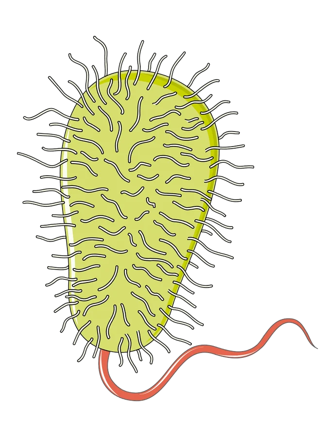
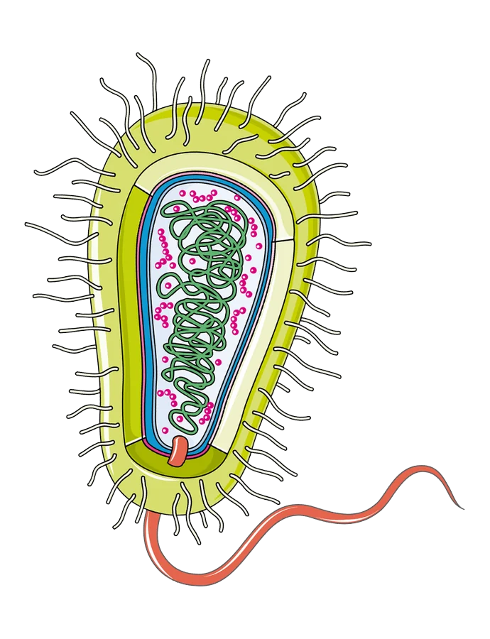
Single-celled organisms (e.g. Amoeba, Euglena) get oxygen and nutrients by diffusion straight through their surface membrane — no circulatory system needed, because their surface area to volume ratio is high enough to supply what they need.
Larger, multicellular animals have a low surface area to volume ratio — their volume (and therefore demand) grows faster than their surface area (supply) can keep up. So they need a circulatory system to actively transport substances to and from cells: oxygen, glucose, carbon dioxide, urea, hormones, and heat.
A bacterium: diffusion is enough
A human: blood carries substances
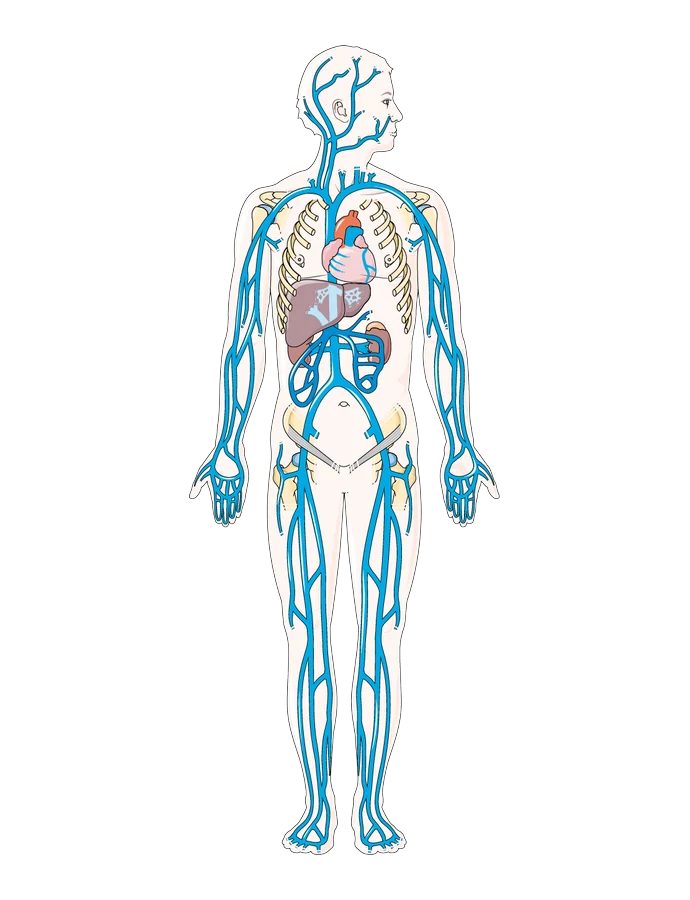
The heart pumps blood out to the head, arms and legs.
2. Open vs closed, single vs double circulation
Closed circulatory system: blood stays inside vessels (e.g. fish, mammals).
Open circulatory system: blood is pumped out into the body cavity and flows freely round the organs, not kept inside vessels (e.g. insects).
Single circulation (e.g. fish): blood passes through the heart once per full circuit of the body — heart → gills → body → heart.
Double circulation (e.g. mammals, including humans): blood passes through the heart twice per full circuit:
- Pulmonary circulation: heart → lungs → heart (picks up oxygen, drops off CO2)
- Systemic circulation: heart → rest of body → heart (delivers oxygen, collects CO2)
Why double circulation is better for active animals:
- Blood pressure is boosted again after the lungs, so it arrives at body tissues faster and at higher pressure than a single circulation could manage.
- Higher flow rate = more oxygen delivered for aerobic respiration, and waste removed faster.
- The septum keeps oxygenated and deoxygenated blood separate on the two sides of the heart, so tissues receive fully oxygenated blood.
A fish: single circulation
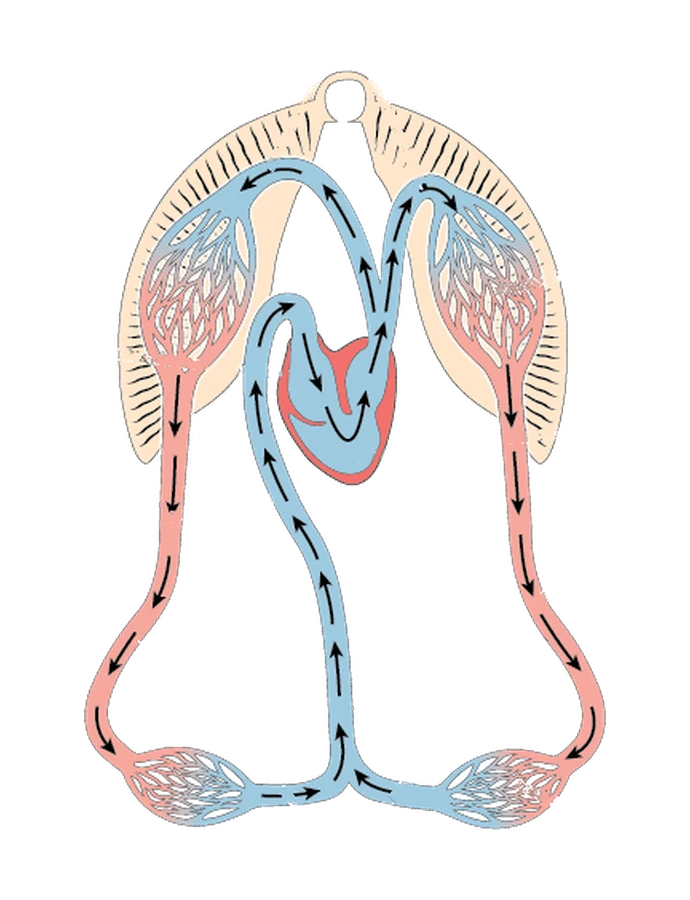
Blood passes through the heart once on each circuit: heart → gills → body → heart.
A human: double circulation
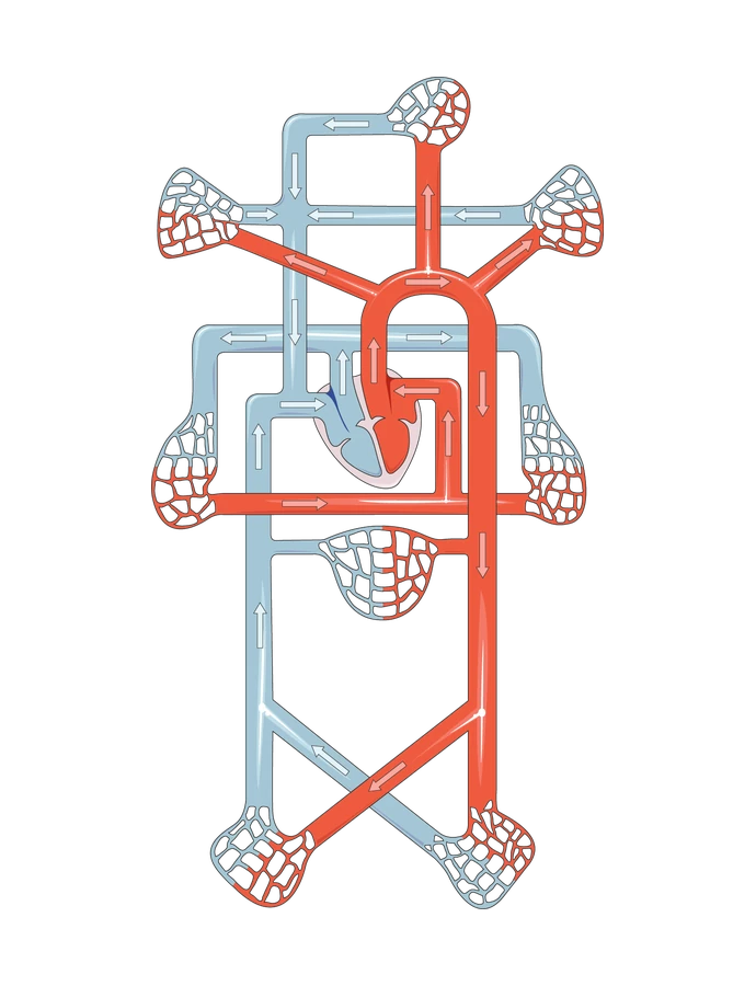
Blood passes through the heart twice on each circuit: heart → lungs → heart → body → heart.
3. The heart’s structure
Four chambers:
| Chamber | Position | Function |
|---|---|---|
| Right atrium | Upper right | Receives deoxygenated blood from the body (via vena cava) |
| Right ventricle | Lower right | Pumps deoxygenated blood to the lungs (via pulmonary artery) |
| Left atrium | Upper left | Receives oxygenated blood from the lungs (via pulmonary vein) |
| Left ventricle | Lower left | Pumps oxygenated blood to the whole body (via aorta) |
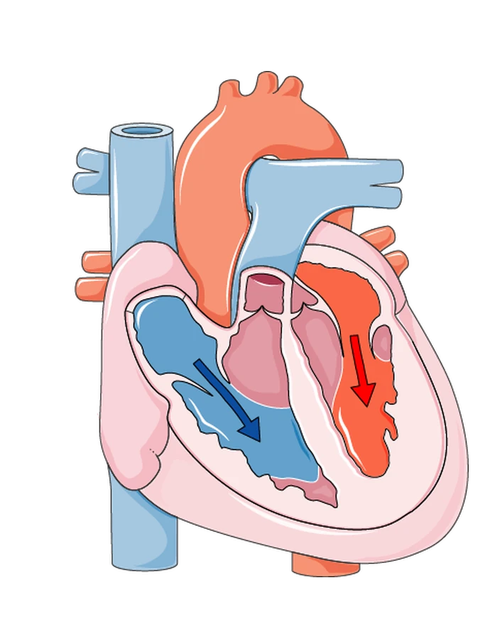
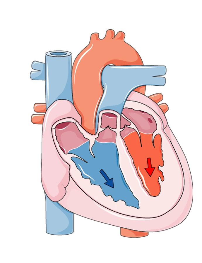
The heart is shown from the front, as if you are facing the person. That is why right and left seem flipped: the heart’s right side is on the left of the diagram.
Slide to see inside the heart
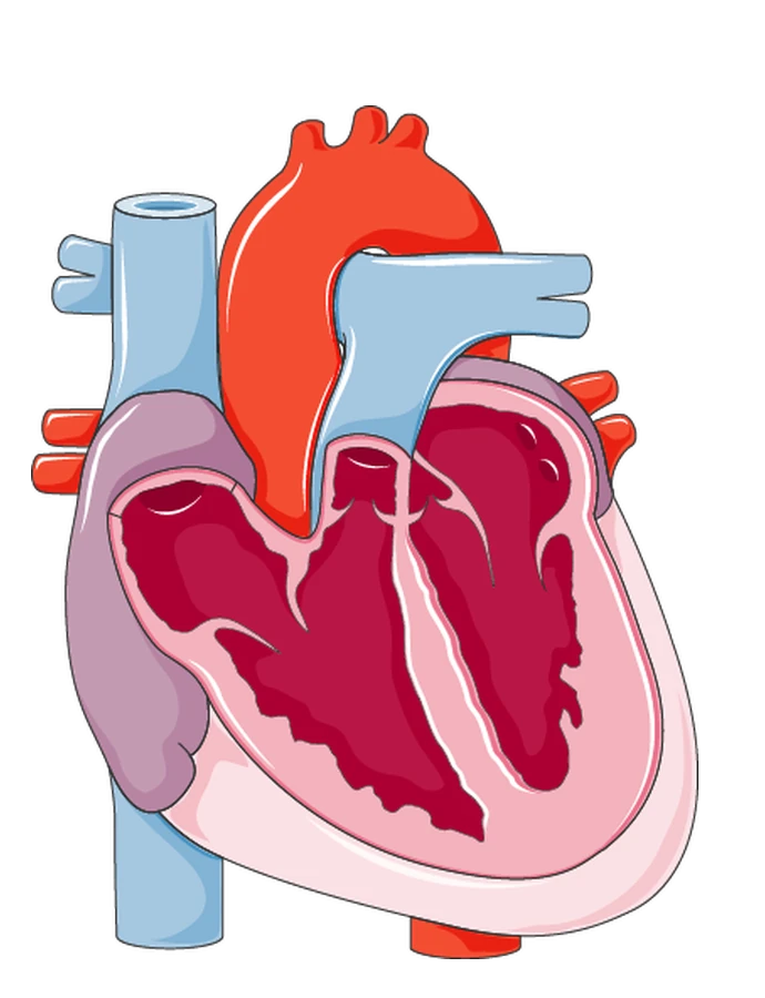
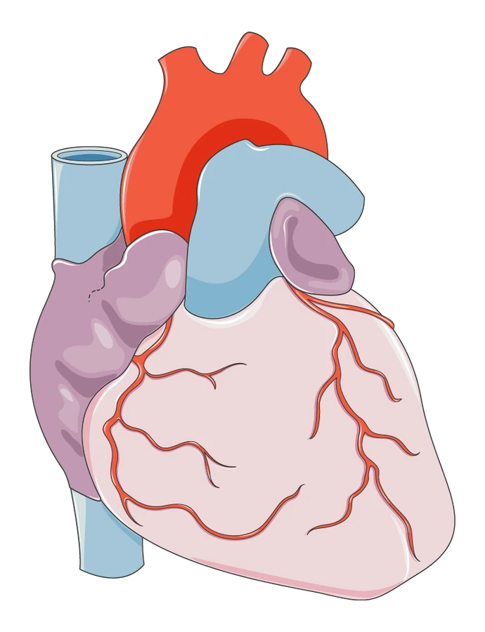
Red: oxygenated blood Blue: deoxygenated blood
Key structural points:
- Atria have thin walls — they only need to push blood a short distance into the ventricles below.
- Ventricles have thick, muscular walls — they push blood much further.
- Left ventricle wall is thicker than the right — it must generate enough pressure to pump blood all around the body, while the right ventricle only pumps to the nearby lungs.
- A septum (wall of muscle) separates the left and right sides, keeping oxygenated and deoxygenated blood from mixing.
- Valves (atrioventricular valves between atria/ventricles; semilunar valves at the base of the aorta and pulmonary artery) prevent backflow, forcing blood to move one way only.
- Cardiac muscle is highly resistant to fatigue — it contracts continuously without tiring, unlike skeletal muscle.
- The pacemaker (a group of cells in the wall of the right atrium) generates the electrical impulses that trigger contraction and sets heart rate (~70 bpm at rest).
- Coronary arteries branch off the aorta and supply the heart muscle itself with oxygenated blood, since the heart is a working muscle with its own respiration needs.
4. The cardiac cycle
One heartbeat, in four stages. Use the arrows to step through them.
-
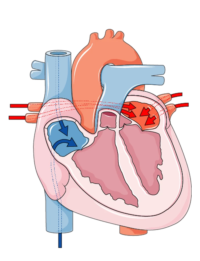
Stage 1 of 4 The atria fill
Deoxygenated blood from the body flows into the right atrium through the vena cava. At the same time, oxygenated blood from the lungs flows into the left atrium through the pulmonary veins.
-
Stage 2 of 4 The atria contract
The atria contract and push blood down through the open tricuspid valve (right side) and bicuspid valve (left side). Deoxygenated blood enters the right ventricle; oxygenated blood enters the left ventricle.
-
Stage 3 of 4 The ventricles fill
The atria empty and both ventricles fill with blood. As the ventricles start to contract, the tricuspid and bicuspid valves close, so blood cannot flow back into the atria.
-
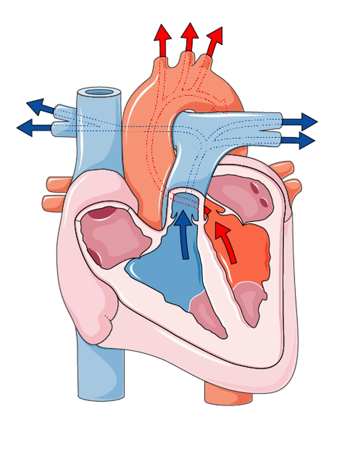
Stage 4 of 4 The ventricles contract
The ventricles contract and the semilunar valves open. Deoxygenated blood leaves the right ventricle through the pulmonary artery to the lungs; oxygenated blood leaves the left ventricle through the aorta to the rest of the body. The cycle then starts again.
5. Blood vessels
| Arteries | Veins | Capillaries | |
|---|---|---|---|
| Direction | Carry blood away from the heart | Carry blood back to the heart | Link arteries to veins through tissues |
| Pressure | High pressure, pulsing | Low pressure, smooth flow | Pressure falls, pulse disappears |
| Wall | Thick, muscular, elastic | Thinner, less muscle/elastic tissue | Wall one cell thick |
| Lumen | Small/narrow | Large | Very narrow (RBCs squeeze through single-file) |
| Valves | None | Yes (prevent backflow) | None |
| Oxygen content | Oxygenated (except pulmonary artery) | Deoxygenated (except pulmonary vein) | Loses oxygen along its length |
Why each is adapted this way:
- Arteries: thick elastic/muscular walls stretch and recoil to withstand and smooth out the high-pressure surges from the ventricles.
- Veins: blood has lost most of its pressure by this point, so thinner walls are fine; valves are essential to stop blood flowing backward, especially against gravity from the lower body; the large lumen (the hollow central cavity inside a blood vessel through which blood flows) reduces resistance to help blood move despite low pressure.
- Capillaries: walls just one cell thick, providing the shortest possible diffusion distance for oxygen, glucose, CO2, and waste to exchange with cells. Networks are huge (billions of capillaries) to maximise surface area for exchange.
Named vessels to know:
- Aorta — main artery, left ventricle → body (highest pressure)
- Vena cava — main vein, body → right atrium
- Pulmonary artery — right ventricle → lungs (only artery carrying deoxygenated blood)
- Pulmonary vein — lungs → left atrium (only vein carrying oxygenated blood)
Arteries
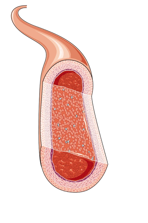
Cross-section
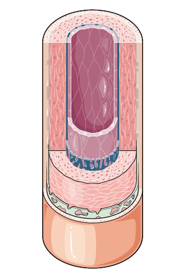
Tunics: the layers that make up the artery wall
Veins
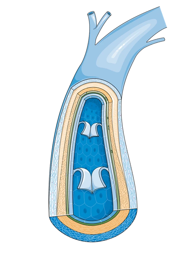
Cross-section
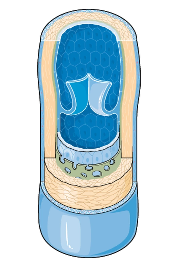
Tunics: the layers that make up the vein wall
Capillaries
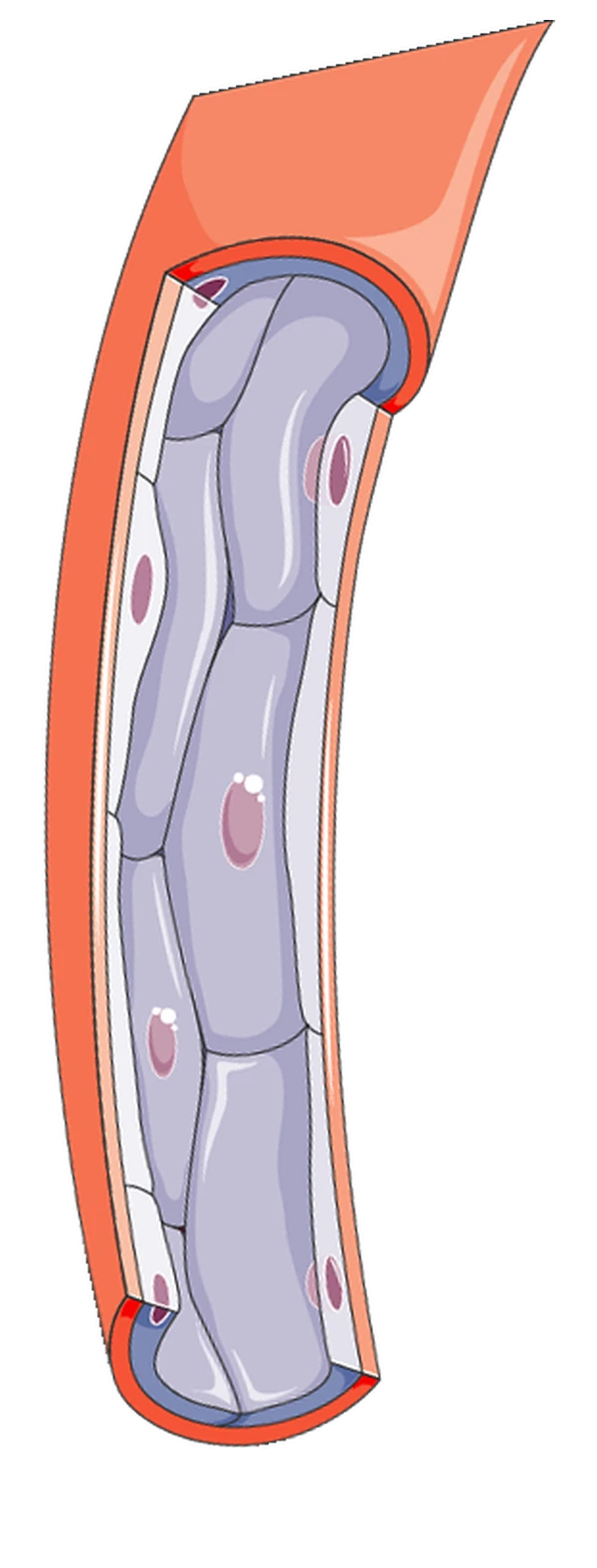
Cross-section
From artery to capillary
-
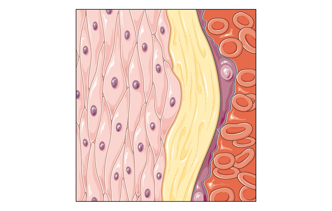
1 of 2 The artery wall
Arteries branch into smaller vessels called arterioles, which act as control valves to regulate blood flow and pressure.
-
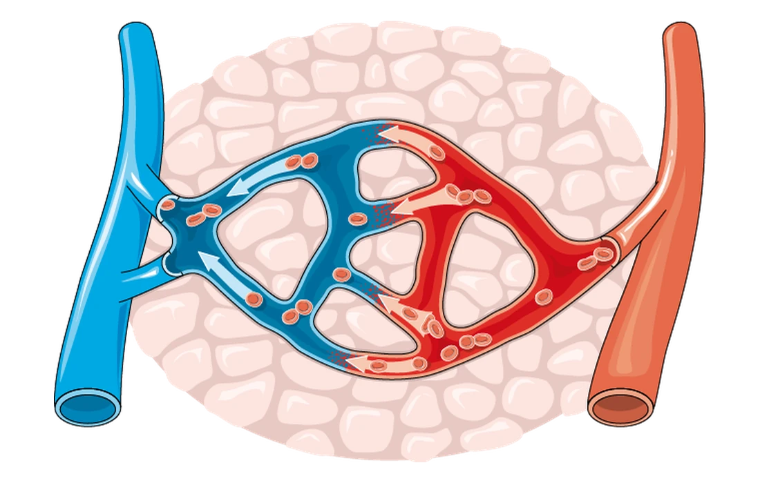
2 of 2 A capillary bed
Blood arrives from an arteriole (right) and flows through a network of capillaries, whose walls are one cell thick. Oxygen and glucose diffuse out of the capillaries into the surrounding cells. Carbon dioxide diffuses from the cells into the capillaries and is carried away towards a vein (left).
Download
Print these and work through them away from the screen.
All of this is free
Every GCSE and FSMQ biology resource on this site is free, with no account needed. Taught sessions are currently GCSE maths only — if you would like a group opened for this subject, join the waiting list and it helps me decide what to run next.
Join the waiting list →